Describe the Structure and Function of the Lungs and Pleura
Further they subdivide into lobes and segments. These two pleural layers resist being pulled apart and therefore attach the lungs to the wall of the thoracic cavity.

Lungs Diagram How Do We Breathe Lungs And Pleura Interactive Biology By Leslie Lung Anatomy Respiratory System Anatomy Human Lungs
Pleural - wraps around each individual lung 3.

. The pleural fluid acts as a lubricant allowing the parietal and visceral pleura to glide over each other friction free. 1 Describe the structure of pleural sac. Both of these layers are completely attached to the surface of the lungs and from t.
The outer surface of the pleural membrane is called the visceral pleura and the membrane that lines the body wall is called the parietal pleura. There are two pleura the parietal and the visceral. Pleura ploorə key membranous lining of the upper body cavity and covering for the lungs.
The pleurae consist of two layers. The outer or parietal pleura lines the inside of the rib cage and the diaphragm while the inner visceral or pulmonary layer covers the lungs. Occupy most of the thoracic cavity.
There are two pleurae in the body. This is a condition of the development of cancer cells in the pleura. Describe the development adult anatomy and function of the lungs and pleural sacs Describe the development adult anatomy and function of the lungs and pleural sacs Introduction Pleural sac is also known as pleural mesothelioma.
Apex of lungs is. With the thoracic cavity the outer pleural membrane is in very close contact. View the full answer.
This fluid is produced by the pleural layers themselves. Lungs have a spongy texture and have a pinkish-gray hue. The function of the pleura is to allow optimal expansion and contraction of the lungs during breathing.
The lungs are enclosed by the double-layered pleural sac. And with the lungs surface the inner pleural membrane is in close contact. Visceral pleura and the parietal pleura.
It contains a small film of. They consist of a serous membrane a layer of simple squamous cells supported by connective tissue. The lung parenchyma also is covered by a pleura123 The purpose of the lung is to provide oxygen to the blood.
The parietal pleura lines the inner surface of the thoracic cavity and ribcage. Each is a double membrane. The pleural membrane is made up of two layers.
The parietal pleura lines the walls of the chest cage and covers the upper surface of the diaphragm and the pulmonary pleura or visceral layer tightly covers the surface of the lungs. Parietal pleura lines the pulmonary cavities. Describe the structure of the lungs and pleural cavities Pleural cavities are found between two layers of a serous membrane.
1 Each of the pleura of the lung is invaginated inorder to form a pleural sac which is primarily composed of two layers ie. The pleura is a two-layered structure. This fluid is secreted by the pleural membrane.
The left lung has a cardiac notch where the heart apex makes an indentation therefore l lung smaller than r. Pleura and pleural membrane are the two membranes that help in enclosing the lungs. Pleural fluid is the fluid that is present in the fluid cavity.
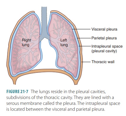
The diaphragm is the flat dome-shaped muscle located at the base of the lungs and thoracic cavity. Each pleural cavity is defined by a space surrounding each lung and is lined by a pleural membrane. The lungs are pyramid-shaped paired organs that are connected to the trachea by the right and left bronchi.
The pleura secrete a fluid that fills the pleural space between the lungs and ribcage to reduce the friction created by the movement of the lungs during inhalation and exhalation. Parietal - all of the thoracic cavity and diaphragm 2. Each pleura can be divided into two parts.
Each lung is contained within a pleural cavity. Turn CO2 blood to O2 blood. This arrangement is very similar to that of the pericardial cavity around the heart Pleural fluid lubricates lungs as they expand and contract to reduce friction Helps prevent lungs from collapsing when you take a breath.
Visceral pleura forms the outermost layer of the lungs. Visceral pleura covers the lungs. They serve as a cushion for the lungs and secrete a fluid that allows them to move easily within the chest cavity.
The visceral pleura line the lungs. The right and left pleurae which enclose the right and left lungs respectively are separated by the mediastinum. The outer surfaces of the lungs are covered with a membrane called the pleura.
The pleura is a sac in the human body which. The lung itself is not located within the pleural cavity rather it is surrounded by it. Pleural cavity lies between the visceral and parietal pleura.
In between the two layers of the pleural membranes there is pleural liquid. On the inferior surface the lungs are bordered by the diaphragm. Each lung is enclosed within a cavity that is surrounded by the pleura.
Pleurae - 3 layers. Lines the walls of the thoracic cavity. The pleura plural pleurae is a serous membrane that surrounds the lung.
This simple squamous epithelial layer is also known as the mesothelium. A pleura is a smooth membrane folded over itself to create a two-layered membrane with a space in. The right lung has three lobes superior middle and inferior where the left lung only has two superior and inferior.
The lungs are enclosed by the pleurae which are attached to the mediastinum. The pleural membrane is thin moist slippery and has two layers. One associated with each lung.
The two pleural membranes surround the lungs which rest on the diaphragm. Also they are anatomically described as having an apex three borders and three surfaces. Pleura of the Lungs.
The Pleural Cavities And Pleural Membranes Structure And Function Organization Of The Respiratory System

22 2 The Lungs Anatomy Physiology
Pin On Lung Cancer
No comments for "Describe the Structure and Function of the Lungs and Pleura"
Post a Comment